As fast and simple as you want it to be
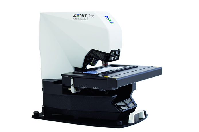
Zenit fast is the new A. Menarini Diagnostics automated system for the acquisition, analysis and storage of IFA slides, which provides laboratories with an efficient solution to improve the management of IFA tests, standardize the validation procedure and reduce intra- and inter-laboratory variability.
What distinguishes Zenit fast is the image autofocusing technology, whole well scanning, the software algorithms for IFA detection and pattern recognition, run-time, types of ANA IFA patterns recognized and its ability to analyze different IFA substrates.

Main features and benefits
- Fast and efficient screening of negative and positive samples
- Fast slide scanning: <30 seconds per well (HEp-2 slides, 20x), >2.5 mins per well for tissues
- Multirun sessions to process large sample numbers
- 8-slide tray
- High-quality scanning sensor and LED microscope illumination
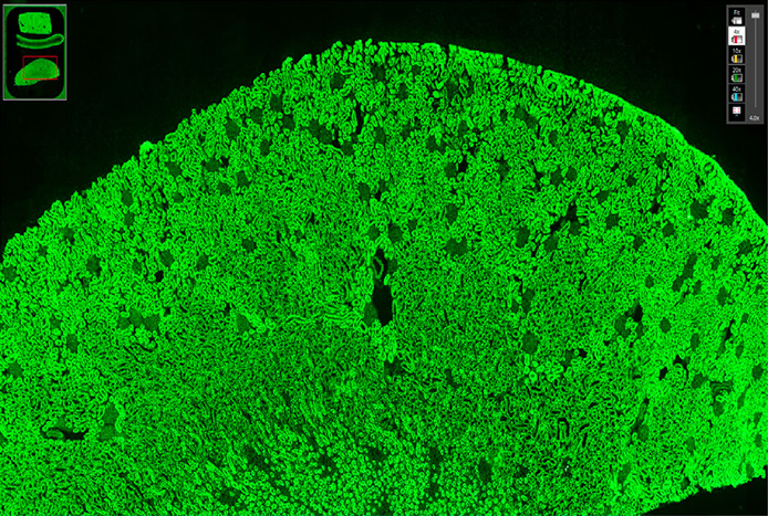
- Whole well scanning
- Virtual microscope for well navigation
- Reduced operating costs

Powerful and reliable information management
Zenit fast offers powerful analytics with a simple and intuitive software interface.
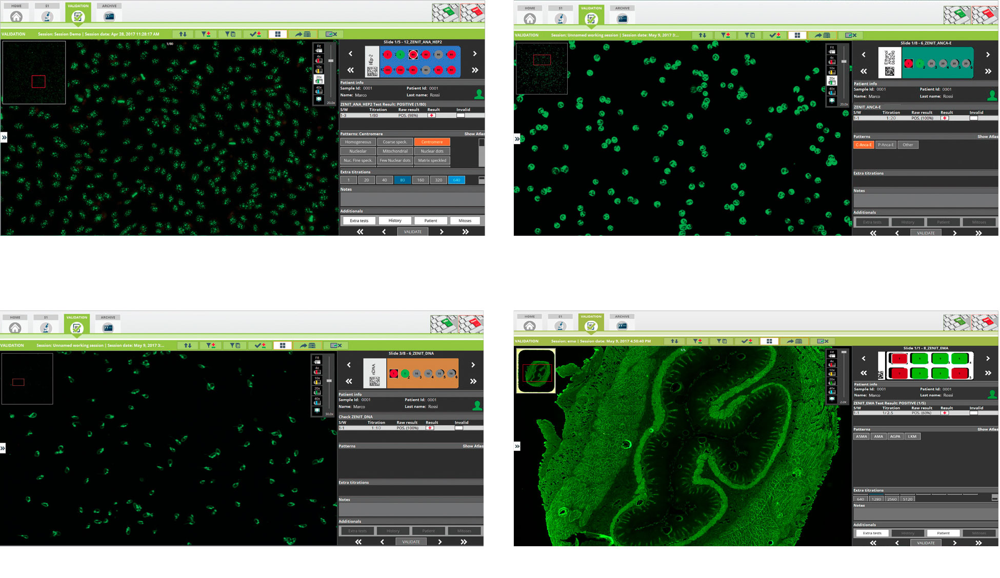
The software is structured into 3 main modules: Scanning, Validation, and Archive.
- Scanning: The user can upload a worklist and launch the automatic scanning of slides that are already mounted and ready on the microscope stage. Scanning can be launched in manual or automatic mode.
- Validation: The digitized image is displayed and can be navigated with the virtual microscope tool. Results are interpreted and validated by the operator using the software interface.
Customizable views facilitate navigation. Results can be sent directly to LIS as soon as each well is validated.
- Archive: The user can access the patient database, performed tests and processed slides. Free text search or multiple and complex queries can be set.
Automated image analysis software
The system performs automatic classification of positive/negative results for Zenit ANA HEp-2 tests measuring the intensity of fluorescence for each positive test and provides pattern recognition on positive results (see detected patterns in the Technical Data section). Mitosis recognition is also performed on identified patterns.
The system is now also capable of discriminating between positive and negative samples for both ANCA E and ANCA F and to define the type of pattern (see detected patterns in the Technical Data section).
A new feature in the software allows also the discrimination between positive/negative results on DNA Crithidia slides.
